 Inverity
Neural CT Compression: 25% Fewer Bits Than JPEG-2000
Author
Brandon Cade
Date Published
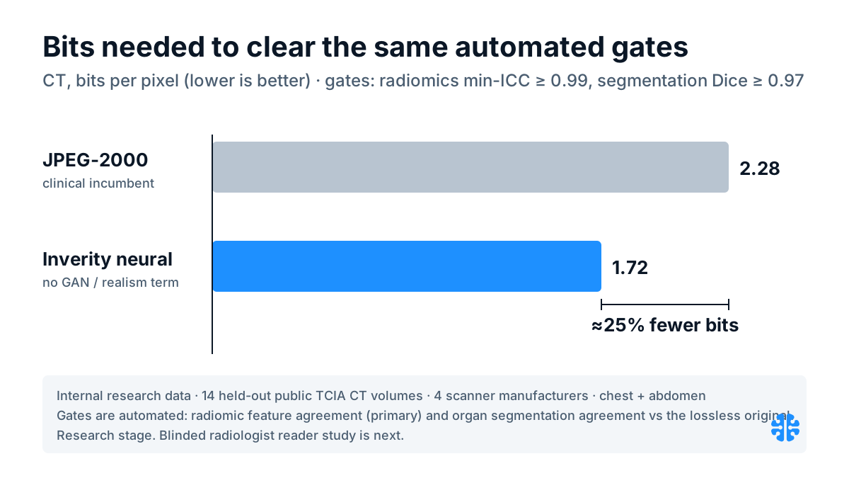
TL;DR. Inverity's neural CT codec cleared the same automated quality gates as JPEG-2000 at 1.72 bits per pixel, versus 2.28 for JPEG-2000. That's about 25% fewer bits, measured on 14 held-out CT volumes from four scanner manufacturers (internal research, July 2026). The codec is trained with no realism term, so it's never rewarded for generating detail. A blinded radiologist reader study is next.
Every bit a codec saves pays off twice: once in the archive, and again each time a study moves between sites or to the cloud. JPEG-2000 is a long-established lossy codec in DICOM. We built a neural medical image compression codec specifically for CT to find out whether it could beat JPEG-2000 while clearing the same automated quality checks that clinical research relies on.
How much smaller is neural medical image compression than JPEG-2000?
About 25% smaller than JPEG-2000 at the same automated quality bar. Our neural codec cleared the pre-specified quality gates at 1.72 bits per pixel. JPEG-2000 needed 2.28 bits per pixel to clear the same gates on the same scans. Put the other way, JPEG-2000 files are roughly 1.33× the size.
| JPEG-2000 | Inverity neural codec |
|---|---|---|
Bits per pixel | 2.28 | 1.72 |
Radiomics gate (min ICC ≥ 0.99) | Cleared | Cleared (0.993) |
Segmentation gate (Dice ≥ 0.97) | Cleared | Cleared (0.976) |
We quote the comparison point where JPEG-2000 clears both gates, because it's the stricter, like-for-like test. The evaluation set was 14 held-out CT volumes from The Cancer Imaging Archive (Clark et al., Journal of Digital Imaging, 2013). It spans four manufacturers, multiple slice thicknesses, chest and abdominal anatomy, and full-dose and low-dose protocols. None of these scans were used in training.
How did we measure image quality?
We compared each compressed scan with its lossless original on two automated tasks that clinical research already relies on. Radiomics is the primary gate. We extracted intensity and texture (GLCM) features from both versions and measured agreement with the intraclass correlation coefficient (ICC). The weakest feature reached an ICC of 0.993, above the 0.99 threshold.
Segmentation is the second gate. TotalSegmentator (Wasserthal et al., Radiology: Artificial Intelligence, 2023) segmented organs on both versions, and we compared the masks. Mean Dice was 0.976 against a 0.97 threshold. That result comes from the segmenter's fast mode. An accurate-mode re-run, which reduces the segmenter's own variability, is part of the next phase.
Why doesn't the codec invent detail?
The codec is trained only to match the original. Many neural image codecs add a realism (GAN) term so their output looks sharper and more natural. That's the right trade for photos and the wrong one for a CT scan. Our medical training objective is pixel error plus bit cost, with the realism weight set to zero. The model is penalized for any difference from the original and never rewarded for plausible-looking texture.
For workflows that need the original exactly, the system also has a byte-exact lossless mode, verified by hash on every operation.
Rules set in advance, metrics audited
Every study uses a pass/fail rule committed to version control before any training or evaluation, so thresholds can't drift after the fact. Held-out sets go through an automated leakage check, and all data is de-identified public data.
We also audit the measurement tools as hard as the model. Three times during the program, a metric looked wrong. Each time we traced it to a flaw in how we were measuring, not to the codec, and fixed it before reporting anything. The model checkpoint never changed.
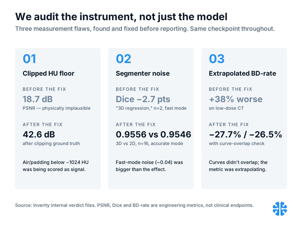
- Ground-truth clipping. One dataset stores air and padding below −1024 Hounsfield units. Clipping the reference to the diagnostic range moved PSNR from an implausible 18.7 dB to 42.6 dB.
- Segmenter variability. An early two-volume test suggested our 3D mode hurt segmentation. On 16 volumes in accurate mode, 3D and 2D were equivalent (0.9556 vs 0.9546 Dice).
- Rate-curve overlap. A low-dose comparison once showed the codec 38% worse. With an overlap check, it was 27.7% and 26.5% cheaper than JPEG-2000.
Does neural medical image compression work on MRI?
The MRI results point the same way. A codec adapted from the CT model achieved a 34% bit-rate reduction versus JPEG-2000 across T1, T2, and FLAIR brain MRI from two manufacturers, on six held-out volumes. On a paired radiomics measure, the difference in feature distortion versus JPEG-2000 stayed within a pre-specified 0.02 equivalence tolerance (bootstrap 95% CI). Next, we'll move the segmentation gate to SynthSeg, a contrast-agnostic segmenter suited to T2 and FLAIR.
Next: a blinded reader study
Radiologists will read compressed and lossless CT side by side, without knowing which is which. The planned design:
- At least 30 cases, sized by a power calculation
- At least two radiologists reading each case
- Randomized crossover reads at the 1.72 bpp operating point
- A pre-registered concordance threshold
- Chest and abdominal CT at minimum
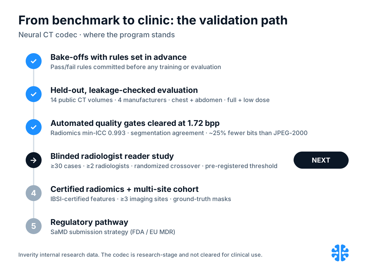
Alongside it, we'll move radiomics to an IBSI-certified implementation (Zwanenburg et al., Radiology, 2020). We'll also extend the cohort across multiple imaging sites and add radiologist-drawn ground-truth masks. Reader studies are how radiology has set lossy compression guidance before, as in the national Canadian evaluation of irreversible compression ratios (Koff et al., Journal of Digital Imaging, 2008). We're following the same path.
Frequently asked questions
Is the codec available for clinical use?
Not yet. The codec is research-stage and hasn't received FDA clearance or CE marking. Compression software intended for diagnostic reads is generally regulated as software as a medical device (U.S. FDA). The blinded reader study is the next step toward that pathway.
Why compare against JPEG-2000?
JPEG-2000 is a long-established lossy codec in DICOM, which makes it the fair baseline. Beating the incumbent is the comparison that matters to hospitals and imaging vendors. Benchmarking against a weaker baseline would inflate the numbers without telling buyers anything useful.
What data was the codec tested on?
Fourteen held-out CT volumes from public, de-identified TCIA collections, spanning four scanner manufacturers. The set covers chest and abdominal anatomy, multiple slice thicknesses, and both full-dose and low-dose protocols. No protected health information was involved, and an automated check confirmed that no evaluation scan appeared in training.
Can I take part in the reader study?
Yes. We're looking for radiologists with chest or abdominal CT experience, and for research sites with independent acquisition chains. If you'd like to read cases or host part of the study, contact Inverity.
Results are Inverity internal research data (CT: July 2026, 14 held-out volumes; MRI: July 2026, 6 held-out volumes) on public de-identified TCIA data. Quality gates are automated measures (radiomics and segmentation agreement). The codec is research-stage and not cleared for clinical use.
Brandon Cade is the founder of Inverity, which builds neural compression for imaging and video.
Sources
- Clark K, et al. "The Cancer Imaging Archive (TCIA): Maintaining and Operating a Public Information Repository." Journal of Digital Imaging, 2013. https://doi.org/10.1007/s10278-013-9622-7 (retrieved 2026-09-30)
- Wasserthal J, et al. "TotalSegmentator: Robust Segmentation of 104 Anatomic Structures in CT Images." Radiology: Artificial Intelligence, 2023. https://doi.org/10.1148/ryai.230024 (retrieved 2026-09-30)
- Zwanenburg A, et al. "The Image Biomarker Standardization Initiative: Standardized Quantitative Radiomics for High-Throughput Image-based Phenotyping." Radiology, 2020. https://doi.org/10.1148/radiol.2020191145 (retrieved 2026-09-30)
- Koff D, et al. "Pan-Canadian Evaluation of Irreversible Compression Ratios ('Lossy' Compression) for Development of National Guidelines." Journal of Digital Imaging, 2008. https://doi.org/10.1007/s10278-008-9139-7 (retrieved 2026-09-30)
- U.S. Food and Drug Administration. "Software as a Medical Device (SaMD)." https://www.fda.gov/medical-devices/digital-health-center-excellence/software-medical-device-samd (retrieved 2026-09-30)